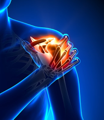
The shoulder is considered as the most complex joint. Therefore, the elements contributing to the stability of the shoulder are no less intricate. Recurrent Shoulder Instability is the persistent inability of the ligaments, tendons and the muscles around the shoulder bone to keep the arm centered in the shoulder socket.
The shoulder is a complex anatomical structure made up of 3 bones:
• Upper Arm or Humerus
• Shoulder Blade or Scapula
• Collar Bone or Clavicle
It is a ball and socket joint that allows multidirectional mobility of hand. The Humerus Head fit into a shallow socket in your shoulder bone called Glenoid. Stability of the shoulder is established by a special harmony between Humerus Head, Glenoid, Labrum, Glenohumeral Ligaments, Rotatory cuffs, Deltoid muscles, and other soft tissue components.
The three main causes of Recurrent Shoulder Instability are listed below:
A careful evaluation and a clear understanding of the patient’s history is the primary step of treatment plan. The Recurrent Shoulder Instability correlates to the type of injury, age at the time of the first dislocation, duration, degree, and direction of instability, hyperlaxity, activity leading to recurrence of dislocation etc. Imaging tests such as X-Ray, MRI Scan, Scapular Y View Image are the primary radiographic evaluations to identify the problem. While X-Ray identifies the injury to the bones, MRI Scan provides a detailed image of soft tissues which helps in identifying the injuries to ligaments and tendons surrounding the shoulder joint.
Depending upon the patient’s condition the doctor will develop a treatment plan to relieve your symptoms. Doctors recommend both surgical and non- surgical treatment plans for patients.
Arthroscopy is a minimal invasive surgery in which soft tissues are repaired through a small incision. During this procedure, an instrument called Arthroscope with a light source and video camera is inserted through a cut into the affected area which provides the surgeon a clear picture to analyze the problem. The surgeon can sometimes correct the problem simultaneously with an additional thin surgical instrument inserted through an additional incision.
In some cases, doctors opt for open surgery. This involves larger incision that requires more healing time.
After the recovery period, patients are directed to do therapies or exercises to strengthen the ligaments and improve the range of motion in their shoulder. Your commitment to these therapies and exercises play an important role in reviving the strength and mobility of your shoulder.
Dr. Biren Nadkarni, Senior Orthopedic and Joint Replacement Surgeon with 15 years of unparalleled service, provides high-quality patient care with ardent dedication and commitment. Currently working in Sitaram Bhartia Institute of Science and Research, New Delhi, Dr. Biren Nadkarni is rendering his distinguished service to Holy Family hospital and Cygnus Orthocare Hospital in New Delhi.
Visit @ www.drbirennadkarni.com
Mail us: drbirennadkarni@gmail.com
Book appointment: www.drbirennadkarni.com/book-appointment.html
Shoulder Anatomy:
The shoulder is a complex anatomical structure made up of 3 bones:
• Upper Arm or Humerus
• Shoulder Blade or Scapula
• Collar Bone or Clavicle
It is a ball and socket joint that allows multidirectional mobility of hand. The Humerus Head fit into a shallow socket in your shoulder bone called Glenoid. Stability of the shoulder is established by a special harmony between Humerus Head, Glenoid, Labrum, Glenohumeral Ligaments, Rotatory cuffs, Deltoid muscles, and other soft tissue components.
Causes:
The three main causes of Recurrent Shoulder Instability are listed below:
- Severe Injury or Trauma:
- Repetitive Strain:
- Multidirectional Instability:
Clinical Evaluation:
A careful evaluation and a clear understanding of the patient’s history is the primary step of treatment plan. The Recurrent Shoulder Instability correlates to the type of injury, age at the time of the first dislocation, duration, degree, and direction of instability, hyperlaxity, activity leading to recurrence of dislocation etc. Imaging tests such as X-Ray, MRI Scan, Scapular Y View Image are the primary radiographic evaluations to identify the problem. While X-Ray identifies the injury to the bones, MRI Scan provides a detailed image of soft tissues which helps in identifying the injuries to ligaments and tendons surrounding the shoulder joint.
Treatment Options:
Depending upon the patient’s condition the doctor will develop a treatment plan to relieve your symptoms. Doctors recommend both surgical and non- surgical treatment plans for patients.
- Non-Surgical Treatments:
- Surgical Treatments:
Arthroscopy:
Arthroscopy is a minimal invasive surgery in which soft tissues are repaired through a small incision. During this procedure, an instrument called Arthroscope with a light source and video camera is inserted through a cut into the affected area which provides the surgeon a clear picture to analyze the problem. The surgeon can sometimes correct the problem simultaneously with an additional thin surgical instrument inserted through an additional incision.
Open Surgery:
In some cases, doctors opt for open surgery. This involves larger incision that requires more healing time.
Recovery:
After the recovery period, patients are directed to do therapies or exercises to strengthen the ligaments and improve the range of motion in their shoulder. Your commitment to these therapies and exercises play an important role in reviving the strength and mobility of your shoulder.
Shoulder Pain Treatment in India
Dr. Biren Nadkarni, Senior Orthopedic and Joint Replacement Surgeon with 15 years of unparalleled service, provides high-quality patient care with ardent dedication and commitment. Currently working in Sitaram Bhartia Institute of Science and Research, New Delhi, Dr. Biren Nadkarni is rendering his distinguished service to Holy Family hospital and Cygnus Orthocare Hospital in New Delhi.
Visit @ www.drbirennadkarni.com
Mail us: drbirennadkarni@gmail.com
Book appointment: www.drbirennadkarni.com/book-appointment.html


